586 Selected Applications
θ
Delay
Object
Plane of
holographic
recording
Scatterer
B
A
D
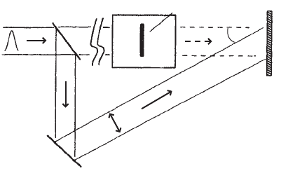
Figure 13.4 Light-in-flight holography illustrated for a simple one-dimensional transparent object
(uniform in the transverse dimension of the object).
sufficiently small angle for the fringes to be resolved by the camera), and recon-
structed through numerical fast Fourier transform [27–29]. The advantage of the
electronic recording and numerical processing is the possibility to integrate a
large number of successive (reconstructed) images. The speckle pattern is aver-
aged out if the time interval between exposures is greater than the correlation
time of the speckle.
13.1.5. Microscopy
Laser-scanning microscopy (see, for example, Wilson and Sheppard [30]) is
ideally suited to combine microscopic imaging with fs pulse illumination. There
are several attractive application fields of femtosecond microscopy—(a) nonlin-
ear microscopy, (b) microscopy with simultaneous space and time resolution,
and (c) microscopy of structures immersed in a scattering environment. An early
review can be found in Kempe and Rudolph [19]. In nonlinear microscopy the
image signal is generated by a nonlinear optical process, such as surface SHG
and two-photon excited fluorescence. An image is a map of the distribution of the
corresponding nonlinear susceptibility. Multiphoton fluorescence is particularly
attractive for microscopy of biological cells because of the depth selectivity of
the excitation process. Since its invention in 1990 by Denk et al. [31] the two-
photon fluorescence microscope has greatly improved the microscopic imaging
capabilities, in particular in the life sciences. Femtosecond pulses are needed
because of their great peak power at comparatively small pulse energy (i.e.,
small heat consumption in the specimen). An overview of ultrafast optics for
biological imaging can be found in a review by Squier [32].

Imaging 587
Simultaneous µm spatial and temporal resolution is of great desire for the
inspection of ultrafast opto-electronic circuits. Another direction is to com-
bine techniques of ultrafast spectroscopy with spatial resolution—to monitor,
for example, diffusion and relaxation of excited carriers in semiconductors.
In fluorescence microscopy additional information can be gained by measur-
ing the lifetime of the fluorescence. Because this relaxation depends sensitively
on the interaction of the fluorescing dye with the environment, the lifetime image
can describe local field and ion concentrations in cells [33].
Another example where scanning can be complemented by temporal correla-
tion with a reference pulse is confocal imaging of objects buried under scattering
layers. Confocal microscopy distinguishes itself by its depth selectivity [see
Figure 13.5]. Enhanced depth selectivity and optical amplification of the image
signal are desirable for imaging through scattering layers that strongly attenuate
the ballistic light. Both aspects can be addressed with scanning microscopy based
on a sensitive correlation technique, such as heterodyning [34]. A realization of
such a microscope is shown in Figure 13.6. It consists of a Michelson interfer-
ometer which contains a scanning microscope in one arm and a piezoelectric
transducer for Doppler shifting the reference pulse in the other arm. The role
of the pinhole is played by the coherent overlap of the plane reference wave and
the image light. A maximum heterodyne signal is obtained if the wave front from
the object is plane (parallel to the reference wave front), that is, if the object is
in focus.
Assuming a layer of thickness L with scattering coefficient µ
s
ontopofan
object with reflectivity R, the image signal of the correlation microscope can be
Beamsplitter
Beamsplitter
Object
Object
Pinhole
Pinhole
Lens
Lens
Object in focus
Object out of focus
PMT
z
PMT
Figure 13.5 Schematic diagram of (reflection) confocal microscopy. Only light from layers that
are in focus can pass through the pinhole and reach the detector. The beam (or object) is scanned
in transverse direction to obtain a two-dimensional image of the layer that can be displayed by a
computer.
Get Ultrashort Laser Pulse Phenomena, 2nd Edition now with the O’Reilly learning platform.
O’Reilly members experience books, live events, courses curated by job role, and more from O’Reilly and nearly 200 top publishers.

