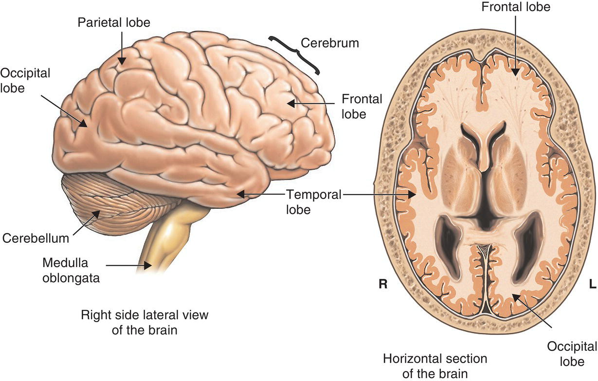
Plate 1 The gross anatomy of the human brain. The image on the left depicts the brain as seen from the side (if the eyes were present, they would be located on the right-hand side of this image); in the image on the right, the brain is viewed from above, as it would look if the top part of the skull and brain had been sliced off.
Source: Nucleus Medical Art/Visuals Unlimited/Science Photo Library.

Plate 2 Photograph of a healthy human brain cut into two halves. This brain has been sliced down the sagittal plane, into its left and right halves (imagine a straight line running from the nose to the back of the head, and slicing along this line). The cut reveals some of the brain's inner anatomy, including the corpus callosum and the third ventricle.
Source: Geoff Tompkinson/Science Photo Library.

Plate 3 The limbic system. An artist's depiction of the limbic system, a network of brain regions that is particularly important for emotional functioning.
Source: 3D4Medical.com/Science Photo Library.

Plate 4 A human neuron. An artist's depiction of a typical human neuron with the main anatomical ...
Get Great Myths of the Brain now with the O’Reilly learning platform.
O’Reilly members experience books, live events, courses curated by job role, and more from O’Reilly and nearly 200 top publishers.

